


Bengaluru: Indian scientists have mapped the brain’s last frontier by creating a high-resolution atlas of the brainstem, offering a clearer view of one of the most vital yet least understood parts of the human brain.
Researchers at the Singapore-India collaborative centre have developed a three-dimensional brainstem atlas using microscope images. The model has identified more than 200 clusters of brain cells and nerve pathways. The structure has been built using 8 chemical markers that distinguish cell types with clarity.
The brainstem remains a small but vital region. It connects the brain to the spinal cord. It controls breathing, heartbeat, sleep, and movement. Damage to small clusters within this area can cause severe consequences. Its dense structure has long limited detailed mapping. This has made it one of the most challenging areas of the brain to study in detail.
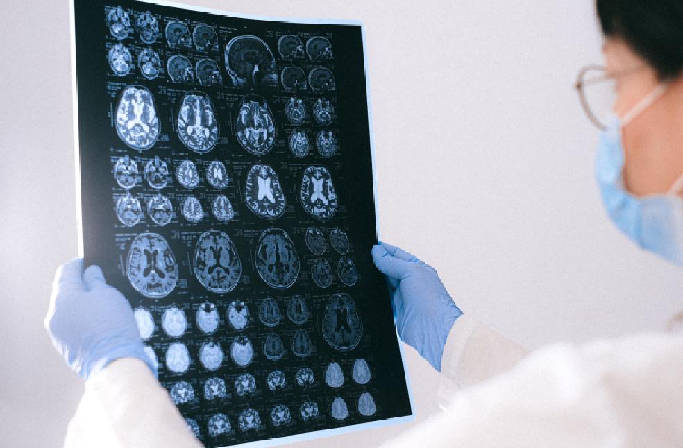
The Singapore Brain Connectivity Group (SGBC) has combined MRI imaging with cellular-level pathology. This approach has allowed users to zoom from full brain scans down to individual neurons. The atlas maintains spatial relationships across scales. This bridges a long-standing gap between imaging and microscopic analysis.
The atlas has been made freely available for global researchers. Scientists have indicated that it could support studies on Alzheimer’s disease, Parkinson’s disease, stroke, and sudden infant death syndrome. Comparisons between healthy and diseased tissue may improve understanding of how disorders develop.
The project has involved around 20 scientists working over 18 months. More than 200 brain sections have been analysed and reconstructed into a digital model. SGBC has stated that future plans include mapping over 100 human brains across different ages and conditions. This effort may help reveal how diseases reshape the brain at the cellular level.
ALSO READ | Taiwan initiates evacuations as Typhoon Bavi hits Japan